
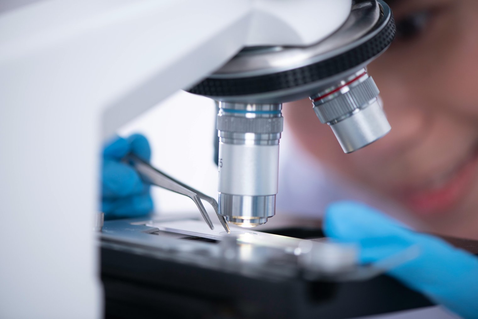
Precision and patience – preparing for a detailed view at the microscopic level.
Preparing a sample for electron microscopy (EM) is a delicate and meticulous process that involves preserving, sectioning, and coating the sample in a way that makes it suitable for examination under an electron microscope. The process allows for extremely high-resolution imaging, revealing structures at the cellular and subcellular level. Proper sample preparation is essential for achieving accurate and high-quality results. It is commonly used in fields like biology, materials science, and nanotechnology.
9:00 AM: The technician gathers necessary materials: biological sample, tweezers, gloves, sample holder, sputter coater.
9:15 AM: The sample is placed into a chemical fixative to preserve its structure, with care to avoid contamination.
9:30 AM: The sample is dehydrated through a series of alcohols to remove water, preventing damage during the process.
9:45 AM: The sample is embedded in a resin for sectioning and placed under a microtome for ultra-thin slicing.
10:00 AM: After slicing, the sample is mounted on a grid and coated with a conductive layer (such as gold) using a sputter coater.
Overview: A premier research facility housing some of the most advanced electron microscopes.:
Landmarks: Scanning electron microscope (SEM), transmission electron microscope (TEM), sample preparation area.:
Tips: Take the time to inspect the sample under a light microscope before proceeding to the electron microscope.:
MIT Research Laboratory of Electronics (Cambridge, MA)
Overview: A world-renowned lab with cutting-edge electron microscopy tools for nanotechnology and materials science research.:
Landmarks: Cleanroom for sample preparation, TEM, SEM.:
Tips: Ensure that all preparation steps are done in a cleanroom to prevent contamination of the sample.:
University of Oxford Materials Science Department (Oxford, UK)
Overview: Houses electron microscopy facilities for studying materials at the atomic level.:
Landmarks: Electron microscope suite, ultra-thin sectioning area.:
Tips: Familiarize yourself with the specific protocols used for materials science sample preparation.:
University of Cambridge Department of Materials Science (Cambridge, UK)
Stanford University Nanofabrication Facility (Stanford, CA)
California Institute of Technology (Caltech) Microscopy Facility (Pasadena, CA)
University of California, Los Angeles (UCLA) Imaging and Microscopy Core (Los Angeles, CA)
Max Planck Institute for Intelligent Systems (Stuttgart, Germany)
Tokyo Institute of Technology Microscopy Lab (Tokyo, Japan)
Swiss Federal Laboratories for Materials Science and Technology (Dübendorf, Switzerland)
Institute of Materials Research and Engineering (Singapore)
Stanford Synchrotron Radiation Lightsource (Stanford, CA)
Harvard University Electron Microscopy Facility (Cambridge, MA)
Tokyo University of the Arts Microscopy Lab (Tokyo, Japan)
University of Minnesota Electron Microscopy Facility (Minneapolis, MN)
Lawrence Berkeley National Laboratory (Berkeley, CA)
University of Chicago Nanotechnology Laboratory (Chicago, IL)
University of Pennsylvania Electron Microscopy (Philadelphia, PA)
ETH Zurich Laboratory of Materials and Nanotechnology (Zurich, Switzerland)
University of Paris-Saclay Microscopy Laboratory (Paris, France)
Arizona State University Microscopy Core Facility (Tempe, AZ)
University of California, San Diego (UCSD) Nanoscience Microscopy Facility (San Diego, CA)
University of Texas at Austin Electron Microscopy Facility (Austin, TX)
University of Tokyo Nanotechnology Research Center (Tokyo, Japan)
University of Edinburgh Advanced Microscopy (Edinburgh, UK)
University of Würzburg Electron Microscopy Center (Würzburg, Germany)
Imperial College London Materials Science Lab (London, UK)
Research Institute of Materials Science (Kyushu University, Japan)
University of Sydney Electron Microscopy Facility (Sydney, Australia)
Precision, meticulousness, scientific exploration, cutting-edge technology, research preparation.
1. Thermo Fisher Scientific: Electron microscopes and sample preparation tools
2. Leica Microsystems: Microscopy equipment
3. Zeiss: Electron microscope components and services
4. Hitachi High-Tech: Electron microscopy products
5. JEOL Ltd: Microscope manufacturer
6. FEI Company: Electron microscopy solutions
7. Carl Zeiss AG: Microscopy and imaging technology
8. VWR International: Lab equipment for electron microscopy
9. Dako: Laboratory chemicals and reagents
10. Entegris: High-purity products for scientific research
11. Amazon: Electron microscopy supplies and accessories
12. B&H Photo Video: Camera equipment for documenting microscopy
13. Grainger: Microscopy parts and accessories
14. McMaster-Carr: Microscopy supplies
15. Fisher Scientific: Reagents for specimen preparation
16. Sigma-Aldrich: Chemical supplier for scientific applications
17. Bio-Rad Laboratories: Electron microscopy equipment and kits
18. EMD Millipore: Chemicals and laboratory products
19. Epson: Projectors for lab presentations
20. LabX: Buy and sell used scientific equipment
21. Science Exchange: Collaborative lab services
1. Amazon: Purchase electron microscopy supplies, chemicals, and equipment.
2. B&H Photo Video: Get a camera setup for documenting electron microscopy results.
3. REI: Buy gloves and lab coats for handling delicate samples.
4. Grainger: Order fine-tuned tweezers and other precision tools for sample handling.
5. McMaster-Carr: Source special sample holders and related microscopy accessories.
6. Target: Get cleaning supplies for preparing your workstation.
7. Home Depot: Set up an ergonomic workstation for long hours of microscopy.
8. Fisher Scientific: Order a high-quality sample preparation kit.
9. Walmart: Buy resin and fixative chemicals for preserving samples.
10. LabX: Find used electron microscopes or related equipment.
• Microscope slide holder: To securely place the sample for the electron microscope.
• Sputter coater: For applying a conductive coating to non-conductive samples.
• Sample preparation kit: Includes necessary tools for cutting, mounting, and preserving the sample.
• Tweezers: For precise handling of small samples.
• Laboratory gloves: To prevent contamination of the sample.
• Fixative solution: To preserve the specimen’s structure.
• Cryoprotectant solution: For biological samples that require freezing.
• Dehydrating agents (e.g., ethanol): To remove moisture from the sample.
• Sputter coater: To ensure the sample’s surface is conductive for electron microscopy.
• Lab notebook: To document the preparation process and results.
• JEOL JEM-2100 (Transmission electron microscope)
• Leica EM ACE600 (Sputter coater)
• Thermo Fisher Scientific Krios (Cryo-electron microscope)
• Hitachi SU5000 (Scanning electron microscope)
• FEI Quanta 200 (Environmental scanning electron microscope)
Sample preparation can be time-consuming.
Precision is critical, and small mistakes can lead to inaccurate results.
Expensive equipment and materials are required for sample preparation.
Some biological samples may degrade during preparation.
Fine adjustments may be needed when switching between different magnifications.
Handling small samples requires extreme care and patience.
Expensive operational costs for maintaining high-end microscopes.
Regular maintenance of the microscope and sample preparation tools.
Accurate documentation of the preparation process.
Careful handling of fragile samples to prevent contamination.
Routine calibration of the microscope for consistent results.
Use of cleanroom protocols to avoid contamination.
Ensuring proper sample storage before and after preparation.
Thorough cleaning of equipment after each use.
Return the sample to its storage if not immediately used.
Finalize notes and document all steps for reproducibility.
Clean the workspace and dispose of any waste materials.
Maintain the microscope and related tools for future use.
Transition to the next phase of research or analysis.
Events
You must log in to add an event.
Events for this Scenario
No events found for this scenario yet.
Experiences
Please log in to share your experience.